The scapular notch: a Uruguayan cadaveric study of 62 dry scapulae
DOI:
https://doi.org/10.55005/v3i1.2Keywords:
scapular notch, suprascapular nerve, anatomy, variations, injury, entrapmentAbstract
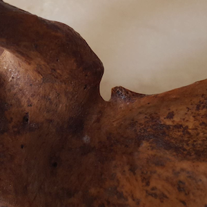
Introduction: The scapular notch is a depression on the superior border of the scapula, located medially to the coracoid process, through which suprascapular nerve enters the supraspinous fossa.
This paper aims to describe the main anatomical aspects of scapular notch, measuring anatomical parameters for identification of this region during surgical procedures, and compare the obtained data with previous worldwide publications.
Material and methods: Sixty-two dry scapulae of Uruguayan specimens were studied at the Anatomy Laboratory of the Faculty of Medicine, Universidad Centro Latinoamericano de Economía Humana (UCLAEH) in Maldonado, and the Faculty of Medicine, University of the Republic in Montevideo, Uruguay, and analyzed for variations.
Results: Of the 62 studied scapulae, 33 were right sided and 29 left sided. Anatomical variations were found in 19 specimens, which included 5 flattened shape notches (8.1%), and 14 ossified notches (22.6%), from which 4 (6.5%) were complete and 10 (16.1%) were incomplete. Scapular notch is located at an average distance of 66.7 mm (SD: 4.7) medially from the lateral border of the acromion.
Conclusions: Anatomy of the scapular notch is variable. The scapular notch can be located at the junction between the medial two thirds and the lateral one third of the superior scapular border. Anatomical variations of this region play an important role in the development of entrapment neuropathies and in surgical considerations for brachial plexus injuries reconstruction.
References
Basta M, Sanganeria T, Varacallo M. Anatomy, Shoulder and Upper Limb, Suprascapular Nerve. [Updated 2021 Oct 9]. In: StatPearls [Internet]. Treasure Island (FL): StatPearls Publishing; Acces 8/1/2022
Tubbs RS, Nechtman C, D ́Antoni AV, et al. Ossification of the suprascapular ligament: A risk factor for suprascapular nerve compression? Int j Shoulder Surg 2013;7(1):19-22. doi: 10.4103/0973-6042.109882
Polguj M, Rozniecki J, Sibinski M, Grzegorzewski A, Majos A, Topol M. The variable morphology of suprascapular nerve and vessels at suprascapular notch: a proposal for classification and its potential clinical implications. Knee Surg Sports Traumatol Arthrosc 2015;23:1542–1548. doi.org/10.1007/s00167- 014-2937-1
Das S, Suri R, Kapur V. Ossification of superior transverse scapular ligament and its clinical implications. Sultan Qaboos Univ Med J 2007;7(2):157–160.
Martínez F: Neuropatía del nervio supraescapular: reporte de dos casos. Rev Urug Med Int 2018; 2: 38-43. doi: 10.26445/rmu.3.2.5
Martínez F. Technical Note: Spinal-Accessory to suprascapular nerve transfer by posterior approach. Austin Neurosurg Open Access 2017;4(1):1058. 7. Martinez F, Jaume A, Sienra C, Russo A. Anatomía quirurgica de la transferencia nerviosa de espinal accesorio a nervio supraescapular por vía posterior. Neurocirugía/Neurocirurgia (FLANC) 2017;26(1):1-6.
Martinez Benia F. Transferencia nerviosa de accesorio-espinal a supraescapular: comparación de vía anterior versus vía posterior. Presentación de una serie y revisión de la literatura. Rev Arg Neurocir 2022;36(1):13-20.
Polguj MB, Sibinski M, Grzegorzewski A, Waszczykowski MB, Majos A, Topol MB. Morphological and Radiological Study of Ossified Superior Transverse Scapular Ligament as Potential Risk Factor of Suprascapular Nerve Entrapment. BioMed Res Int 2014; doi: 10.1155/2014/613601
Polguj MB, Sibinski M, Grzegorzewski A, Grzelak P, StefaNczyk L, Topol M. Suprascapular Notch Asymmetry: A Study on 311 Patients. BioMed Research Int 2014, Article ID 196896.
Jezierski H, Podgórski M, Wysiadecki G, Olewnik Ł, De Caro R, Macchi V, Polguj M. Morphological Aspects in Ultrasound Visualization of the Suprascapular Notch Region: A Study Based on a New Four-Step Protocol. J Clin Med 2018;7(12):491. doi: 10.3390/jcm7120491
Kumar A, Sharma A, Singh P. Anatomical study of the suprascapular notch: quantitative analysis and clinical considerations for suprascapular nerve entrapment. Singapore Med J 2014;55(1):41-4. doi: 10.11622/smedj.2014009 13. Bayramoğlu A, Demiryürek D, Tüccar E, Erbil M, Aldur MM, Tetik O, Doral MN. Variations in anatomy at the suprascapular notch possibly causing suprascapular nerve entrapment: an anatomical study. Knee Surg Sports Traumatol Arthrosc 2003;11(6):393-8. DOI: 10.1007/s00167-003-0378-3
Sangam MR, Sarada Devi SS, Krupadanam K, Anasuya K. A study on the morphology of the suprascapular notch and its distance from the glenoid cavity. J Clin Diagn Res 2013;7(2):189-92. doi: 10.7860/JCDR/2013/4838.2723
Polguj M, Jędrzejewski K, Majos A, Topol M. Variations in bifid superior transverse scapular ligament as a possible factor of suprascapular entrapment: an anatomical study. International Orthopaedics (SICOT) (2012) 36:2095–2100. doi.org/10.1007/s00264-012-1618-2
Moriggl B, Jax P, Milz S, Büttner A, Benjamin M. Fibrocartilage at the entheses of the suprascapular (superior transverse scapular) ligament of man— a ligament spanning two regions of a single bone. J Anat 2001;199(Pt 5):539- 45. DOI: 10.1046/j.1469-7580.2001.19950539.x
Ohanisian L, Brown N, White SD, Rubay D, Schwartz PM. Persistent Shoulder Pain Due to a Suprascapular Nerve Injury in the Setting of Trauma. Cureus 2019;11(3):e4224. doi: 10.7759/cureus.4224
Daripelli S, Tolupunoori B, Vinodini L. Morphometric Study of Suprascapular Notch and Its Safe Zone in Indian Population. Maedica (Bucur) 2020;15(4):461-467. doi: 10.26574/maedica.2020.15.4.461
LongR,WangN,LiuW,LiuZ,ChengS,ZhangX,GengX,LiuC,WangJ,Ding M, Tian Z, Li J. An anatomical study of the superior transverse scapular ligament of Jining population. Surg Radiol Anat 2019;41(11):1345- 1349. DOI: 10.1007/s00276-019-02330-8
Sinkeet SR, Awori KO, Odula PO, Ogeng’o JA, Mwachaka PM. The suprascapular notch: its morphology and distance from the glenoid cavity in a Kenyan population. Folia Morphol (Warsz) 2010;69(4):241-5.
Polguj M, Jędrzejewski KS, Topol M. Sexual dimorphism of the suprascapular notch – morphometric study. Arch Med Sci 2013;9(1):177-83. DOI: 10.5114/aoms.2013.33173
Yavari M, Hassanpour SE, Alizadeh Otaghvar H, Abdolrazaghi HA, Farhoud AR. The Incidence of Ossified Superior Transverse Scapular Ligament during Nerve Transfer through Posterior Shoulder Approach. Arch Bone Jt Surg 2019;7(3):258-262.
Bhandari PS, Sadhotra LP, Bhargava P, Singh M, Mukherjee MK, Bhatoe HS. Dorsal approach in spinal accessory to suprascapular nerve transfer in brachial plexus injuries: technique details. Indian J Neurotrauma 2010;7(1):71-4. doi: 10.1016/S0973-0508%2810%2980015-2
Colbert SH, Mackinnon S. Posterior approach for double nerve transfer for restoration of shoulder function in upper brachial plexus palsy. Hand (N Y). 2006;1(2):71-7. doi: 10.1007/s11552-006-9004-4
Agrawal D, Singh B, Dixit SG, Ghatak S, Bharadwaj N, Gupta R, Agrawal GA, Nayyar AK. Morphometry and variations of the human suprascapular notch. Morphologie 2015;99(327):132-40. DOI: 10.1016/j.morpho.2015.04.002
Kluver C, Naranja Sotelo M. Ascendencia – Étnica y Seguridad Social en Uruguay. Asesoría en Políticas de Seguridad Social. 2019. Accessed: 12.6.2022: https://www.bps.gub.uy/bps/file/16400/1/65.-ascendencia-etnica-y- seguridad-social-en-uruguay.-c.-kluver-y-m.-naranja-sotelo.pdf
Kannan U, Kannan NS, Anbalagan J, Rao S. Morphometric study of suprascapular notch in Indian dry scapulae with specific reference to the incidence of completely ossified superior transverse scapular ligament. J Clin Diag Res 2014;8(3):7-10. doi: 10.7860/JCDR/2014/7836.4089
Albino P, Carbone S, Candela V, Arceri V, Vestri AR, Gumina S. Morphometry of the suprascapular notch: correlation with scapular dimensions and clinical relevance. BMC Musculoskelet Disord 2013; 14:172. doi.org/10.1186/1471-2474-14-172
Joy A, Sinha MB, Satapathy BC. The Ossified Bifid Superior Transverse Scapular Ligament Causing a Double Suprascapular Foramen: A Case Report. J Clin Diag Res 2015;9(6): AD03-AD04. doi: 10.7860/JCDR/2015/12812.6037
Yang HJ, Gil YC, Jin JD, Ahn SV, Lee HY. Topographical anatomy of the suprascapular nerve and vessels at the suprascapular notch. Clin Anat. 2012;25(3):359-65. DOI: 10.1002/ca.21248
Kharay SS, Sharma A, Singh P. Unusual morphology of scapulae: incidence and dimensions of ossified ligaments and supraspinous bony tunnels for clinical consideration. Singapore Med J 2016;57(1):29-32. doi: 10.11622/smedj.2015103
Bagoji IB, Hadimani GA, Bulgoud RS, Desai V, Prakash KG, Bharatha A. Anatomical Variations of the Suprascapular Notch and its Importance in Suprascapular Entrapment Neuropathy. Maedica (Bucur) 2020;15(3):298-304. doi: 10.26574/maedica.2020.15.3.298
Łabętowicz P, Synder M, Wojciechowski M, Orczyk K, Jezierski H, Topol M, Polguj M. Protective and Predisposing Morphological Factors in Suprascapular Nerve Entrapment Syndrome: A Fundamental Review Based on Recent Observations. Biomed Res Int 2017;:4659761. doi: 10.1155/2017/4659761
Downloads
Published
How to Cite
Issue
Section
License
Copyright (c) 2023 Fernando Martínez, Guzmán Ripoll, Alejandra Neirreiter, Carlos Rodríguez, Karla Duarte

This work is licensed under a Creative Commons Attribution 4.0 International License.
Authors retain copyright of their work, with first publication rights granted to the publisher.


