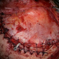
Temporalis muscle reattachment by using transosseus running suture along superior temporal line: technical note
DOI:
https://doi.org/10.55005/v2i1.2Abstract
Introduction: After reattachment of the temporalis muscle, atrophy of the temporalis muscle may occur, which is associated with difficulty in chewing function. To prevent this, numerous surgical modifications have been made to allow reattachment of the temporalis muscle with minimal damage.
Methods: We describe the technical details of surgical modification for reattachment of the temporalis muscle in 12 cases treated surgically in our department.
Results: We used a transosseous continuous suture along the superior temporal line as a base for reattachment of the muscle. The temporalis muscle was successfully reattached in all observed cases. No infections, dislocations, muscle tears, or significant temporal atrophy with depression occurred in any of the observed cases. In the author's technique, the temporalis muscle is reconstructed anatomically at the level of the superior temporal line. At follow-up after approximately 24 months, all patients were satisfied with the cosmetic result.
Conclusions: The use of running sutures along the superior temporal line is a safe, simple, and successful alternative for reattachment of the temporal muscles in patients undergoing surgery for intracranial pathology. The surgery takes slightly longer but does not require additional costs. This technique minimizes the risk of atrophy of the temporal muscles. With this technique, muscle tension was maintained with good stabilization and the cosmetic result is also satisfactory.
References
Oikawa S, Mizuno M, Muraoka S, Kobayashi S. Retrograde dissection of the temporalis muscle preventing muscle atrophy for pterional craniotomy. Technical note. J Neurosurg. 1996;84(2):297-299. doi:10.3171/jns.1996.84.2.0297
Chierici G, Miller AJ. Experimental study of muscle reattachment following surgical detachment. J Oral Maxillofac Surg. 1984;42(8):485-490. doi:10.1016/0278-2391(84)90006-5
Zager EL, DelVecchio DA, Bartlett SP. Temporal muscle microfixation in pterional craniotomies. Technical note. J Neurosurg. 1993;79(6):946-947. doi:10.3171/jns.1993.79.6.0946
Hochberg J, Kaufman H, Ardenghy M. Saving the frontal branch during a low frontoorbital approach. Aesthetic Plast Surg. 1995;19(2):161-163. doi:10.1007/BF00450252
Bowles AP, Jr. Reconstruction of the temporalis muscle for pterional and cranioorbital craniotomies. Surg Neurol. 1999 Nov;52(5):524-9.
Spetzler RF, Lee KS. Reconstruction of the temporalis muscle for the pterional craniotomy. Technical note. J Neurosurg. 1990;73(4):636-637.doi:10.3171/jns.1990.73.4.0636
Stechison MT. Temporal muscle fixation. J Neurosurg. 1995;82(4):701-702.
Wells MD, Kirn DS. Bone anchors: new application in craniofacial surgery. J Craniofac Surg. 1996;7(2):164-167.
Hönig JF. V-tunnel drill system in craniofacial surgery: a new technique for anchoring the detached temporalis muscle. J Craniofac Surg. 1996;7(2):168-169. doi:10.1097/00001665-199603000-00021
Castillo R, Zarate A, Chavez R. Reconstruction of the temporalis muscle. Journal of
Neurosurgery. 1992 Feb;76(2):336. DOI: 10.3171/jns.1992.76.2.0336.
Webster K, Dover MS, Bentley RP. Anchoring the detached temporalis muscle in craniofacial surgery. J Craniomaxillofac Surg. 1999;27(4):211-213. doi:10.1016/s1010-5182(99)80031-6
Eppley BL, Sadove AM, Havlik RJ. Resorbable plate fixation in pediatric craniofacial surgery. Plast Reconstr Surg. 1997;100(1):1-13. doi:10.1097/00006534-199707000-00001
Habal MB. Fixation, imaging, and resorption. J Craniofac Surg. 1996;7(5):325. doi:10.1097/00001665-199609000-00001
Rozema FR, Levendag PC, Bos RR, Boering G, Pennings AJ. Influence of resorbable poly(L-lactide) bone plates and screws on the dose distributions of radiotherapy beams. Int J Oral Maxillofac Surg. 1990;19(6):374-376. doi:10.1016/s0901-5027(05)80086-4
Peltoniemi HH, Tulamo RM, Toivonen T, Hallikainen D, Törmälä P, Waris T. Biodegradable semirigid plate and miniscrew fixation compared with rigid titanium fixation in experimental calvarial osteotomy. J Neurosurg. 1999;90(5):910-917. doi:10.3171/jns.1999.90.5.0910

Downloads
Published
How to Cite
Issue
Section
License
Copyright (c) 2022 Dinko Štimac, Dragan Janković

This work is licensed under a Creative Commons Attribution 4.0 International License.
Authors retain copyright of their work, with first publication rights granted to the publisher.


